
Fine-Tuning Vision After Cataract Surgery
Explains why blur persists, how stability is checked, and when glasses, LAL adjustment, LASIK, PRK, YAG, or lens procedures may fit.
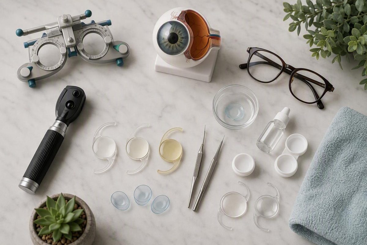
Fine-tuning vision after cataract surgery: options and timing
What “fine-tuning” means—and when blur is urgent instead
Contact your surgical team promptly or seek urgent eye care for sudden or worsening vision loss, significant pain, increasing redness, marked light sensitivity, flashes, many new floaters, double vision, or a curtain-like shadow in your field of vision. These are not symptoms to treat as routine healing or a glasses problem. An ophthalmology provider’s postoperative guidance lists these among warning signs requiring prompt assessment. Review the warning signs described by Southwest Eye Institute.
“Fine-tuning vision after cataract surgery” is not the name of one procedure. It is a cause-first process: identify why vision remains blurry, fluctuating, distorted, uncomfortable, or less glasses-independent than expected, then match the response to that cause.
Cataract surgery removes the cloudy natural lens and replaces it with an artificial intraocular lens, or IOL. Removing the cataract does not guarantee that every part of the visual system will produce the intended result. Early healing, corneal swelling, the tear film, residual prescription, IOL position, capsule clarity, retinal or optic-nerve health, and adaptation to new lens optics can all influence vision.
For that reason, postoperative blur should not automatically be labeled a “refractive miss.” Dry eye calls for a different response from a rotated toric IOL. Posterior capsule clouding requires a different treatment from residual astigmatism. Sudden deterioration may require urgent medical care rather than elective vision correction.
The practical questions are:
- What is limiting the vision?
- Is it improving, stable, fluctuating, or worsening?
- Are the eye and its measurements stable enough to guide treatment?
- What is the least invasive option that addresses the diagnosed cause?
This article is a general cause-and-options overview, not a diagnosis, candidacy assessment, or substitute for professional care. Its procedural evidence comes largely from medical-center and eye-clinic patient education rather than independent comparative guidelines, so it cannot establish which procedure is safest or best for an individual eye. See the site’s Terms of Use for the general-information limitation.
The recovery timeline: healing first, stable measurements second
Mild haze or blur can occur during the first few days after cataract surgery. Many people notice improvement within days, followed by continued sharpening over several weeks. One ophthalmology-practice guide describes final clarity as commonly developing over approximately four to eight weeks, while emphasizing that individual recovery varies. This is a broad pattern—not a deadline, guarantee, or automatic indication for treatment. Greenwich Ophthalmology describes the postoperative recovery pattern and possible causes of persistent blur.
There are two separate timing questions:
- When is the eye stable enough for a useful glasses prescription?
- When is it stable enough for an elective procedure such as LASIK, PRK, or lens-based surgery?
A final refraction—the test used to determine a glasses prescription—is often considered after several weeks, once early healing and swelling have settled. Elective procedural enhancement may be deferred for several months because permanently reshaping the cornea or operating inside the eye requires more confidence that the result is stable.
The calendar alone cannot prove stability. A prescription that changes between visits, ongoing inflammation, significant dryness, prior corneal refractive surgery, irregular astigmatism, or another eye condition may justify waiting or additional testing. Conversely, a medical problem discovered early may require active treatment rather than more observation.
What documented stability means
A clinician may compare findings from more than one visit and ask:
- Is visual acuity improving, unchanged, or worsening?
- Does refraction produce a meaningful improvement?
- Is the measured prescription substantially consistent between visits?
- Is the tear film healthy enough to produce repeatable measurements?
- Has postoperative inflammation settled?
- Is the IOL centered and stable?
- If the IOL is toric, is it properly oriented?
- Are the retina and optic nerve healthy enough to support the expected vision?
- Do the symptoms match the measured refractive error?
Neuroadaptation is different from physical healing
Neuroadaptation is the brain’s process of learning to interpret a changed visual signal. It can be particularly noticeable with multifocal and extended-depth-of-focus IOLs, which distribute light differently from a conventional monofocal lens. Halos, glare, or an unfamiliar balance among near, intermediate, and distance vision may become less intrusive as adaptation occurs.
Neuroadaptation should not become a catch-all explanation, however. Persistent, severe, or worsening glare, halos, blur, or distortion still deserve examination. Dry eye, residual astigmatism, IOL position, capsule clouding, and retinal disease may create similar complaints.
The supplied evidence does not show that special eye exercises, puzzles, visual drills, or brain-training applications accelerate adaptation. Florida Eye Specialists advises using vision normally through activities such as reading, computer use, and television and states that no method has been proved to speed the process. Another commercial clinic recommends training activities but provides no cited clinical studies supporting them. Florida Eye Specialists discusses ordinary visual use and the lack of a proven acceleration method.
Follow your own surgeon’s instructions about drops, eye protection, hygiene, activity, driving, and follow-up. A generic recovery timeline should never replace instructions specific to your operation.
Find the cause before choosing the fix
Symptoms provide clues, not diagnoses. Different conditions can feel similar, and more than one cause may be present at the same time.
| Symptom pattern | What it may suggest | Why examination matters |
|---|---|---|
| Early haze that steadily improves | Routine healing, temporary corneal swelling, or resolving inflammation | Worsening rather than improvement changes the level of concern |
| Blur that varies with blinking or through the day | Tear-film instability or another ocular-surface problem | Residual prescription and other conditions may coexist |
| Burning, grittiness, watering, or irritation with variable focus | Dry eye or ocular-surface disturbance | Medication effects, inflammation, and infection must be distinguished clinically |
| Consistently out-of-focus distance or near vision | Residual nearsightedness, farsightedness, or astigmatism | Refraction alone does not exclude corneal, lens, retinal, or capsule problems |
| Halos or glare with a multifocal or extended-depth-of-focus IOL | Lens optics and neuroadaptation | Persistent symptoms can also arise from dryness, refractive error, PCO, or IOL position |
| Vision that clears and later becomes cloudy again | Posterior capsule opacification may be considered | Retinal and other causes of recurrent blur also require assessment |
| Sudden decline, flashes, many floaters, pain, redness, or a curtain-like shadow | A potentially urgent postoperative or retinal problem | Prompt clinical assessment is needed |
Residual refractive error
A residual refractive error, sometimes called a refractive miss, means that nearsightedness, farsightedness, or astigmatism remains after the implanted lens settles. The eye may be clearer and healthier than before surgery yet still need optical correction for precise focus.
Consistently blurred distance vision, near vision, or both can fit this pattern. So can a difference between the two eyes. Diagnosis requires refraction and examination; symptoms alone cannot establish it.
Dry eye and ocular-surface disturbance
A medical-device manufacturer’s educational article identifies refractive miss, ocular-surface disease, capsule clouding, and optical effects as distinct possible causes requiring different responses; as manufacturer material, it should be read as patient education rather than independent guidance. RxSight outlines these cause-specific categories.
Glare, halos, and optical adaptation
Multifocal and extended-depth-of-focus IOLs are intended to provide a broader range of focus than a conventional single-focus lens. Their optics may also produce halos, glare, or reduced contrast in some settings.
Adaptation may make these effects less noticeable, but waiting indefinitely is not a substitute for evaluation. The assessment may consider the IOL design, residual prescription, pupil characteristics, ocular surface, capsule clarity, lens position, and retinal health.
Cloudiness after an initially clear period
The cataract itself does not grow back because the cloudy natural lens has been removed. The thin capsule left in place to support the IOL can, however, become cloudy or wrinkled. This is posterior capsule opacification, or PCO.
PCO may cause haze, glare, halos, or reduced detail that resembles the original cataract. If examination confirms that PCO is responsible, YAG laser capsulotomy may be considered to create an opening in the cloudy capsule.
Other causes that are not glasses problems
Persistent blur can also arise from inflammation, corneal swelling, cystoid macular edema, IOL displacement, retinal disease, or optic-nerve disease. These problems are not corrected by changing a glasses prescription and may require medical or surgical care.
What the assessment may involve
The examination generally moves from symptoms to measurements:
- History: When did the problem begin? Is it improving, stable, fluctuating, or worsening? Which distances and activities are affected?
- Visual acuity and refraction: How clearly does each eye see, and does a measured prescription improve vision?
- Ocular-surface evaluation: Is the tear film stable? Are there signs of corneal or eyelid-surface disease?
- Corneal measurements: If laser treatment is being considered, testing may assess thickness, shape, regularity, and astigmatism.
- IOL assessment: Is the lens centered and stable? If it is toric, is its orientation contributing to the problem?
- Capsule examination: Is PCO causing haze or glare?
- Retinal and optic-nerve assessment: Can the back of the eye support the expected visual result?
Testing is expanded according to the findings. The objective is to avoid treating the cornea for a problem arising from the tear film, capsule, implanted lens, retina, or optic nerve.
Start with the least invasive cause-specific options
Once urgent and medical causes have been addressed, the next step is often the least invasive option that meets the person’s functional needs.
Stabilize the ocular surface first
If an ocular-surface problem is identified, clinicians may treat it and repeat the measurements before making a permanent refractive decision. Improving the surface may sharpen vision by itself and make refraction or corneal scans more reliable.
Postoperative dry-eye care must be individualized. Recently operated eyes may still require prescribed medicines and surgeon-specific restrictions. Do not stop, replace, or supplement postoperative drops solely because an article recommends artificial tears, preservative changes, warm compresses, or another regimen.
After treatment and further healing, refraction can be repeated. If the result changes substantially, a permanent refractive procedure should not be planned from the earlier measurement.
Glasses
Glasses are the simplest way to compensate for a stable residual prescription. They do not change the cornea or implanted lens and can be updated if visual needs evolve.
They may be prescribed for:
- distance vision;
- reading;
- intermediate work such as desktop screens;
- progressive or multifocal use;
- a specific working distance;
- selected differences between the eyes, when tolerable.
Needing glasses does not by itself mean cataract surgery failed. A monofocal IOL is generally chosen around one principal focal target, so glasses may still be needed at other distances. Toric IOLs address astigmatism but do not guarantee spectacle independence. Multifocal and extended-depth-of-focus designs broaden the intended range but may still leave a residual prescription or task-specific need.
Contact lenses
Contact lenses may be an optical option for suitable patients with a stable prescription.
Contact lenses do not treat inflammation, PCO, IOL displacement, or retinal disease.
Choosing glasses or contacts is not “giving up” on an enhancement. For some people, avoiding another procedure provides the preferred balance of clarity, convenience, risk, and cost.
How a Light Adjustable Lens is refined after implantation
Postoperative light adjustment is available only when a Light Adjustable Lens was implanted during cataract surgery. Ultraviolet treatment cannot convert a conventional fixed-power IOL into an adjustable lens.
A Light Adjustable Lens contains photosensitive material. After implantation and initial healing, a dedicated device delivers targeted ultraviolet light that changes the lens’s shape and focusing power without removing it.
UT Southwestern describes a pathway in which testing and adjustment generally begin about three weeks after surgery. Each light session takes approximately two minutes or less. Up to three adjustments may be performed, often at roughly weekly intervals, followed by two final treatments that lock the lens so it can no longer be adjusted. The medical center describes the adjustment period as commonly lasting four to six weeks, with potentially longer waits after previous LASIK or radial keratotomy. These details describe that center’s reported pathway, not a universal schedule. UT Southwestern explains the adjustment and lock-in process.
This is different from waiting for conventional LASIK, PRK, or lens exchange. Light adjustment is a planned part of care for that specific implant, not a method for modifying any IOL.
UV-protective glasses are a core requirement
UT Southwestern states that patients must wear the prescribed UV-protective glasses during waking hours from immediately after surgery through the final lock-in treatment because the lens remains light-sensitive. Patients should follow the treating team’s exact instructions about eyewear, light treatments, lock-in, and when protection may stop.
The process requires repeated testing and treatment visits. Factors considered during candidacy may include:
- eye anatomy and overall ocular health;
- pupil dilation;
- previous eye surgery;
- visual goals;
- ability to attend repeated appointments;
- ability and willingness to use protective glasses as directed;
- whether the desired distance-near balance is realistically achievable.
That does not establish eligibility or guarantee a particular result.
Neither LAL nor LAL+ should be presented as a promise of 20/20 vision or complete freedom from glasses. The final experience still depends on the cornea, tear film, retina, optic nerve, healing, visual goals, and response to the selected optical target.
LASIK or PRK for a stable residual prescription
LASIK and PRK reshape the cornea. They do not adjust, remove, or replace the implanted IOL.
In selected patients, either procedure may address stable residual nearsightedness, farsightedness, or astigmatism:
- LASIK creates a corneal flap, reshapes tissue beneath it, and repositions the flap.
- PRK treats the corneal surface without creating a LASIK flap and generally involves a slower surface and visual recovery.
Neither procedure is categorically best. Selection depends on the eye’s anatomy and health, prescription, previous procedures, lifestyle, visual goals, and the clinician’s assessment.
Why a glasses prescription is not enough to establish candidacy
A post-cataract laser evaluation may include:
- repeated refraction;
- corneal thickness measurement;
- topography or tomography;
- astigmatism mapping;
- tear-film and dry-eye assessment;
- pupil measurement;
- confirmation of IOL stability;
- retinal examination;
- review of previous LASIK, PRK, radial keratotomy, or corneal disease;
- relevant systemic and inflammatory health history.
A private-clinic overview lists uncontrolled dry eye, thin or irregular corneas, keratoconus or other corneal disease, scarring, unstable refraction, retinal instability, and active autoimmune or inflammatory disease as reasons to avoid treatment or proceed cautiously. These are clinic-described screening considerations, not a substitute for examination or an independent guideline. Eye Clinic London describes its assessment and selection factors.
Timing is individualized. Some clinic sources describe waiting at least three months and sometimes three to six months before laser enhancement, but these are practice patterns rather than universal rules. Stability of refraction, corneal healing, dryness, inflammation, and IOL position matters more than reaching a particular date.
PRK may be considered for selected residual refractive errors when corneal and general eye health permit. An academic medical-center guide likewise emphasizes stable vision and assessment of corneal thickness, refractive-error magnitude, coexisting eye disease, and overall health rather than treating elapsed time as the only criterion. WashU Medicine discusses PRK after cataract surgery.
LASIK and PRK both involve another procedure, follow-up, and recovery. The evidence supplied for this overview is inadequate for comparing complication rates or declaring one option universally safer, more accurate, or better.
One clinic article cites a comparison involving only 65 eyes. Because the underlying study is not available here for critical appraisal, that summary should not be used to promise success rates or rank corneal and lens-based procedures.
When the lens or capsule—not the cornea—needs attention
“Lens adjustment” is too vague for informed decision-making. At least four materially different interventions may be discussed, and they do not treat the same problem.
Toric-IOL rotation
A toric IOL is designed to correct astigmatism at a particular orientation. If it rotates away from the intended axis, its astigmatism correction may be reduced. Toric-IOL rotation repositions the existing lens.
This is not the same as changing an LAL with light, adding another lens, exchanging the implant, or reshaping the cornea. Examination must first confirm that the toric lens’s orientation is contributing meaningfully to the visual problem.
Piggyback IOL
A piggyback procedure places an additional lens in front of the existing IOL to alter the eye’s focusing power. It may enter the discussion when a residual refractive error is not being addressed with corneal laser treatment.
Because it places another implant inside the eye, this is an intraocular procedure—not an optical aid or corneal touch-up. Candidacy and procedure-specific risks require individualized surgical assessment.
IOL exchange
IOL exchange removes the implanted lens and replaces it with another.
Exchange is a more involved, selected intraocular procedure rather than a routine “small adjustment.” A clinic overview distinguishes toric rotation, piggyback implantation, and exchange while also noting that enhancement involves another procedure and recovery period. Its descriptions are useful for defining the options but do not provide independent comparative safety evidence. Blue Fin Vision describes these post-cataract procedures.
YAG laser capsulotomy
Posterior capsule opacification is clouding or wrinkling of the capsule that supports the IOL. The removed natural cataract has not returned.
YAG laser capsulotomy creates an opening in the cloudy posterior capsule so light can pass through more clearly. It treats capsule clouding. It does not correct residual nearsightedness, farsightedness, or astigmatism.
If capsule clouding and a possible lens procedure are both under consideration, ask the surgeon how the sequence should be planned. The supplied evidence does not establish a universal sequence, so the decision must reflect the examination, the degree of PCO, the implanted lens, and the procedures being considered.
The available sources also do not provide robust comparative complication rates or a universal formula for choosing among toric rotation, piggyback implantation, and IOL exchange. Definitions of these procedures should not be mistaken for enough information to select one.
Compare the pathways and prepare for the consultation
The useful question is not, “Which technology is best?” It is, “Which option addresses the diagnosed cause with an acceptable burden, recovery, uncertainty, and set of tradeoffs?”
The table below summarizes roles rather than recommending treatment. Timing and candidacy remain individualized.
| Option | Treats | Changes | Usual stage | Burden and limits | Key uncertainty |
|---|---|---|---|---|---|
| Observation and neuroadaptation | Improving early blur or adaptation to new optics | No anatomy | Early recovery, with monitoring | Follow-up and symptom tracking; inappropriate for urgent or worsening symptoms | No adaptation period applies to everyone |
| Ocular-surface treatment | Dryness or unstable tear film | Tear film and ocular surface | Whenever identified | Treatment varies and must fit the postoperative eye | Improvement and timing vary |
| Glasses | Stable residual prescription | External optics | After refraction becomes reliable | Fitting and possible updates | Does not treat medical, capsule, or IOL-position problems |
| Contact lenses | Stable residual prescription in suitable patients | Optics on the cornea | After clinician clearance | Requires safe handling and adequate surface health | Comfort and suitability are individual |
| LAL light treatment | Residual focus in an implanted LAL | Compatible IOL power and shape | During the implant’s postoperative adjustment pathway | Multiple treatments, lock-in visits, and required UV protection | Applies only to an implanted LAL |
| LASIK | Selected stable refractive error | Cornea beneath a flap | After healing and stability are documented | Procedure, recovery, and corneal candidacy testing | No supplied evidence establishes universal superiority |
| PRK | Selected stable refractive error | Corneal surface | After healing and stability are documented | Slower surface recovery than LASIK in general | Comparative post-cataract evidence is limited |
| Toric-IOL rotation | Reduced astigmatism correction from lens orientation | Position of the existing toric IOL | After malorientation is confirmed | Intraocular procedure and follow-up | Robust comparative rates are not supplied |
| Piggyback IOL | Selected residual focusing error | Adds a second intraocular lens | After stable assessment and planning | Intraocular surgery | Comparative safety and effectiveness remain uncertain |
| IOL exchange | Selected lens mismatch, malposition, or refractive problem | Removes and replaces the IOL | After detailed evaluation | More involved intraocular surgery and recovery | No universal selection threshold is supplied |
| YAG capsulotomy | Clinically meaningful PCO | Opens the cloudy capsule | When PCO is diagnosed | Laser treatment and directed follow-up | Does not correct a residual glasses prescription |
Bring a functional vision record
Before the appointment, note:
- whether vision is improving, unchanged, fluctuating, or worsening;
- whether blinking briefly clears the image;
- whether distance, reading, screens, dashboards, faces, or another task is affected;
- whether one eye is more troublesome;
- glare, halos, starbursts, ghost images, or double vision;
- difficulty with night driving;
- burning, grittiness, watering, or dryness;
- previous LASIK, PRK, radial keratotomy, retinal procedures, or other eye surgery;
- every current eye drop and medicine;
- the implanted IOL type, if known;
- willingness to use glasses or contact lenses;
- ability to attend additional visits or manage another recovery period.
Bring actual working distances when relevant. “Computer vision” may mean a laptop at arm’s length, a large monitor farther away, or detailed work much closer to the face.
Ask about binocular goals
Ask:
- What distance-near balance are we trying to achieve?
- Is one eye intended for a different focal range?
- How does the second eye’s current or planned treatment affect the target?
- Could a temporary optical correction help me understand the proposed balance?
- What tradeoffs could affect depth perception, night vision, or comfort?
The supplied evidence does not establish one planning method for everyone. The appropriate balance depends on daily tasks, current vision in both eyes, implanted lenses, tolerance for imbalance, and willingness to use glasses.
Questions for the surgeon or eye-care clinician
Consider asking:
- What is the most likely cause of my symptoms?
- What examination findings support that explanation?
- Is my refraction repeatable across visits?
- Has the ocular surface been evaluated and stabilized?
- Is the IOL centered, stable, and correctly oriented?
- Is the capsule clear?
- Are the retina or optic nerve limiting vision?
- What happens if I defer treatment?
- Would glasses or contact lenses be a reasonable first option?
- What tests are needed before a laser or lens procedure?
- What recovery, restrictions, and follow-up would apply?
- Which procedure-specific risks matter in my eye?
- Could treating one problem reveal another limitation?
- How will success be defined for my daily activities?
- What result could still require glasses?
Request written cost, insurance, and enhancement-policy details directly from the provider. Coverage and bundled-treatment policies vary, and the supplied evidence does not support general claims about what a clinic or insurer will pay.
A realistic definition of success is better functional vision with an acceptable balance of tradeoffs—not guaranteed 20/20 acuity, perfect vision, or complete freedom from glasses.
The cause-first action plan is:
- Report urgent symptoms immediately.
- Otherwise, track whether vision is improving, fluctuating, consistently defocused, or becoming cloudy again.
- Attend scheduled follow-up examinations.
- Allow healing and address any ocular-surface problem.
- Obtain a reliable, repeatable refraction.
- Compare the least invasive correction with any appropriate procedural option.
The best plan depends on the diagnosed cause, implanted lens, corneal and retinal health, visual priorities, and tolerance for further visits or procedures. Follow-up may involve coordinated care between the surgeon and another qualified eye-care clinician, but examination findings and treatment plans should be communicated clearly across the team.
How long after cataract surgery should I wait before getting new glasses or considering an enhancement?
New glasses are often considered after several weeks, once early healing and the prescription are sufficiently stable. Provider timelines commonly describe stabilization over approximately four to eight weeks, but repeatable measurements matter more than the date.
LASIK, PRK, or a lens-based enhancement may be delayed for several months. Some private clinics describe waiting at least three months and sometimes three to six months, but this is a practice pattern rather than a universal rule. Eye Clinic London describes this timing as dependent on refractive, corneal, surface, and IOL stability.
Can a regular intraocular lens be adjusted with light after cataract surgery?
No. Postoperative ultraviolet-light adjustment works only with a Light Adjustable Lens implanted during cataract surgery. A conventional fixed-power IOL cannot be converted into an adjustable lens with light treatment.
When a conventional IOL leaves a residual prescription, possible approaches may include glasses, contact lenses, selected corneal laser treatment, or a lens-based procedure. The appropriate option depends on the diagnosed cause and examination findings.
Can dry eye make my cataract-surgery result look blurry or fluctuate?
Yes. An unstable tear film can create blur that varies with blinking, time of day, screen use, or environmental conditions. Burning, grittiness, watering, and fluctuating focus can support suspicion of dry eye, but they do not prove the diagnosis.
Dryness should be assessed before relying on a final refraction or corneal scan for permanent treatment. Use postoperative medicines as directed and ask the surgical team before adding, stopping, or replacing eye drops.
Do eye exercises or brain-training apps speed adaptation after cataract surgery?
The supplied evidence does not establish that special visual exercises, puzzles, or brain-training applications accelerate neuroadaptation. Ordinary activities such as reading, watching television, and using a computer may be resumed as allowed by the surgeon, but they should not be presented as proven ways to speed recovery.
Persistent or worsening halos, glare, blur, or distortion should be examined rather than attributed automatically to a need for more “training.”
Can a cataract come back after surgery?
No. The cloudy natural lens was removed, so that cataract cannot regrow.
Vision can become cloudy later if the capsule supporting the IOL becomes cloudy or wrinkled. This is posterior capsule opacification. If examination confirms that PCO is responsible, YAG laser capsulotomy can create an opening in the cloudy capsule. It does not change the IOL prescription or correct residual nearsightedness, farsightedness, or astigmatism.